Technology at The Retina Clinic London
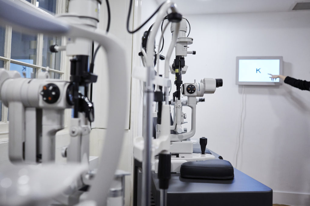
Your consultation with us will be longer than in many other clinics. This is because of the highly-personalised and detailed approach we take. Professor Stanga will complete a full vitreoretinal assessment of your eyes and based on this you will undergo a number of scans which help to diagnose and guide your treatment plan.
Technology we have within the clinic
In addition to industry standard retinal diagnostic and treatment devices, we have access to the latest technology in the field including:
Optos® Ultra-Widefield Retinal Imaging scanner
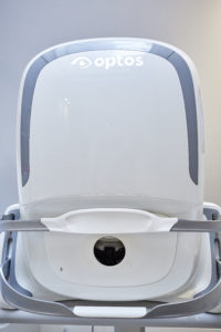 The Optos® Ultra-Widefield Retinal Imaging scanner is able to capture photographs of the retina to a wider degree than any other on the market, up to 200 degrees. Other retinal imaging scanners are restricted to 45 degrees per image, limiting detailed visibility to the central area of the retina and ease of diagnosis.
The Optos® Ultra-Widefield Retinal Imaging scanner uses different light wavelengths (colours) that permit the “layer by layer” visualisation of the back of the eye.
An example of using wavelengths is in the early diagnosis of Dry ARMD whereby short blue light wavelengths is utilised for the diagnostic test known as Fundus Auto-fluorescence.
This imaging technology allows for Ultra-Widefield Fundus Fluorescein Angiography, an excellent tool to assess the retinal blood flow and diagnosis of abnormal blood vessels within the macula region, the central part of the retina, up-to the peripheral retina with high resolution. The use of a dye injected into the vein of your arm can detect conditions such as diabetic retinopathy and vein occlusion. There is no radiation used and the broad retinal views captured during the angiography can enable the early diagnosis, planning and management into your treatment.
Swept-Source DRI Topcon Triton® OCT
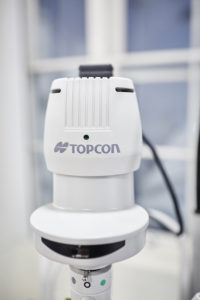 Swept-Source OCT technology combines faster scanning and infra-red light for superior visualisation compared to standard OCT scanners. Infra-red light used is invisible, making the scan more comfortable and less distracting for the patient. This new and advanced imaging technology allows better identification of the outer layer of the vitreous (the clear gel that fills the space between the lens and the retina), the interface between the vitreous and the retina and a greater number of layers within the retina itself as well as under the retina, all with the same sensitivity. This technology allows the visualisation of the intraocular structures in 3D, which makes it easier for the patient to understand the internal architecture of the eye. SS-OCT can take detailed high-quality images which are less affected by eye-movements and cataract formation compared to standard scanners.
This test can confirm or rule out Posterior Vitreous Detachment (PVD) and is an important diagnostic test for patients with reduced or distorted central vision, for example in the presence of an Epiretinal Membrane (ERM) or Macular Hole.
Swept-Source DRI Topcon Triton® OCT also allows the assessment of retinal blood flow and the diagnosis of abnormal blood vessels in the macula, the central part of the retina, in a non-invasive manner without the need of an injection of dye in the vein of your arm. This is an important diagnostic test for Diabetic Maculopathy and Retinopathy and patients with AMD.
This technology aids early detection, management & monitoring of vitreo-retinal conditions.
Topcon Autorefractor
Topcon Autorefractor® can carry out multiple tasks with one simple click on the device. This machine is widely used across optometric practices and UK hospitals. Its purpose is to estimate your prescription, measure your corneal thickness and curvature of the clear front surface of your eye (cornea) non-invasively, it can also measure your intra-ocular pressures with a simple puff of air to each eye. These measurements are routinely conducted to discover any abnormal changes that can relate to glaucoma.
Pascal® Laser
PASCAL® Pattern Scanning Laser technology uses targeted laser pulses to treat the specific area of the retina. PASCAL® technology employs continuous uniform pulses at high-speed to treat the targeted area which is painless and has fewer side effects compared to other laser technologies. This laser technology offers the potential for reduced loss of vision and visual field in the treatment of Diabetic Maculopathy and Retinopathy.
Laser can be delivered into the eye through a Contact Lens or an Indirect Ophthalmoscope, a headscope worn by Prof. Stanga. The Indirect Ophthalmoscope allows treatment of the extreme retinal periphery and Retinal Tears which may not be reachable through a contact lens.
ELLEX® Laser
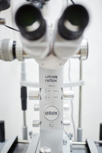
ELLEX® unique Reflex Technology® has been specially engineered to allow clear visualisation deep in the vitreous for the effective treatment of Vitreous Floaters, unlike other YAG laser technologies. This procedure can provide relief from eye floaters impacting vision and causing focusing issues.
 The Retina Clinic London